Our model organ to study organogenesis: The Hydro-vascular organ (HVO)
The sea star hydro-vascular organ (HVO) is a simple structure for buoyancy composed of two parallel tubes that elongate to fuse into a vascular structure.
These epithelial tubes are transparent, not covered by other tissues and can fully regenerate. Like for vertebrate organs, elongation is driven by proliferation and migration and branches are formed using the same sets of genes (Perillo et al., 2023).
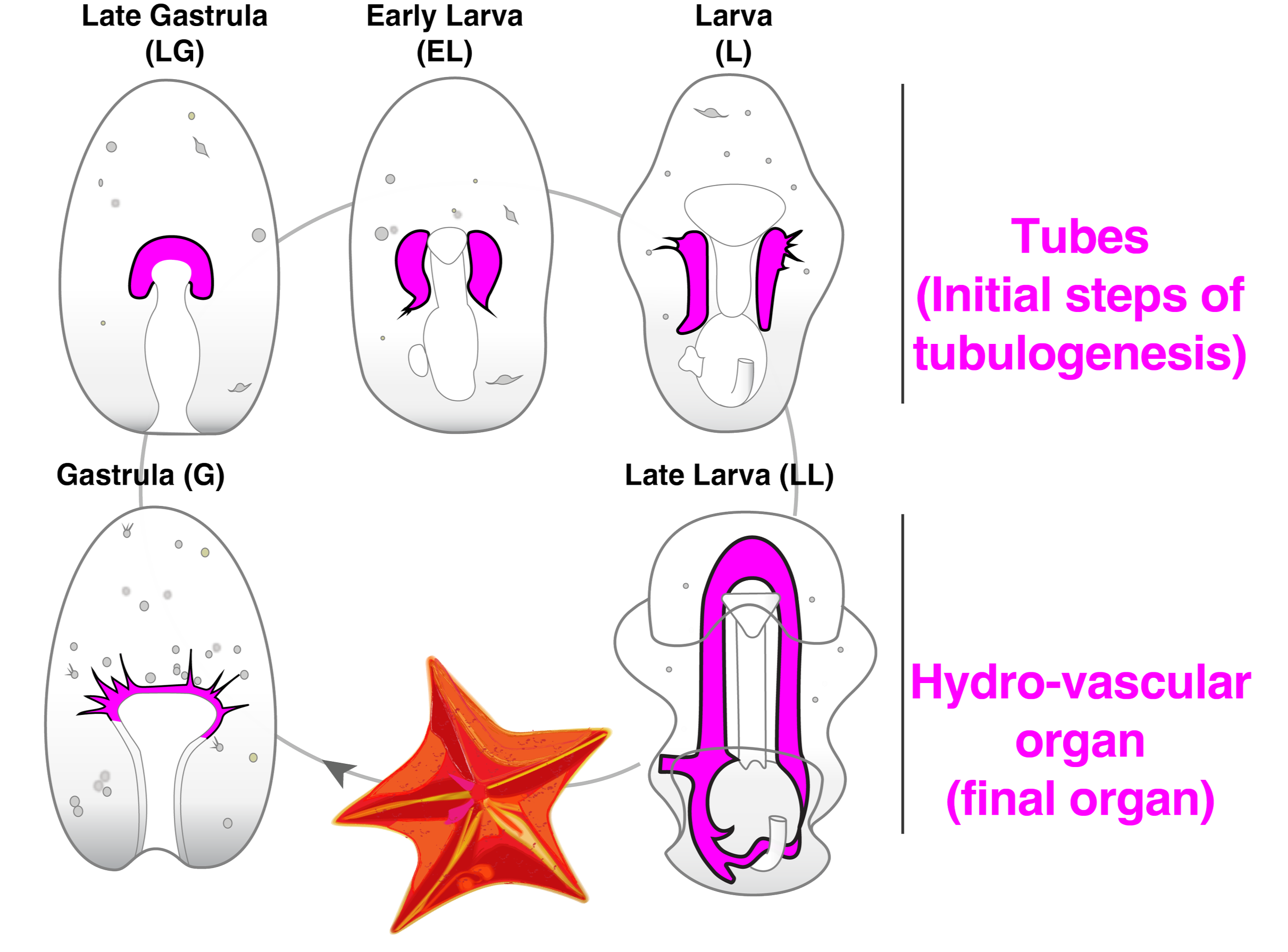
Sea star lifecycle, Hydro-vascular organ in magenta.
Defining the conserved cell types that build tubular organs
All organs grow through the same stages that are finely regulated over time by gene regulatory networks (GRNs) that integrate signaling pathways, transcription factors, and their target genes. What are the nodes in the gene regulatory network that direct morphogenesis of the growing tube? We are generating tissue-enriched single nucleus RNAseq datasets (snRNAseq) to determine how cell lineages change over time from the initial tube to a complex organ.
This project is funded by a NIH/NIGMS MIRA ESI GRANT and partially by a CZI Billion Cells project.
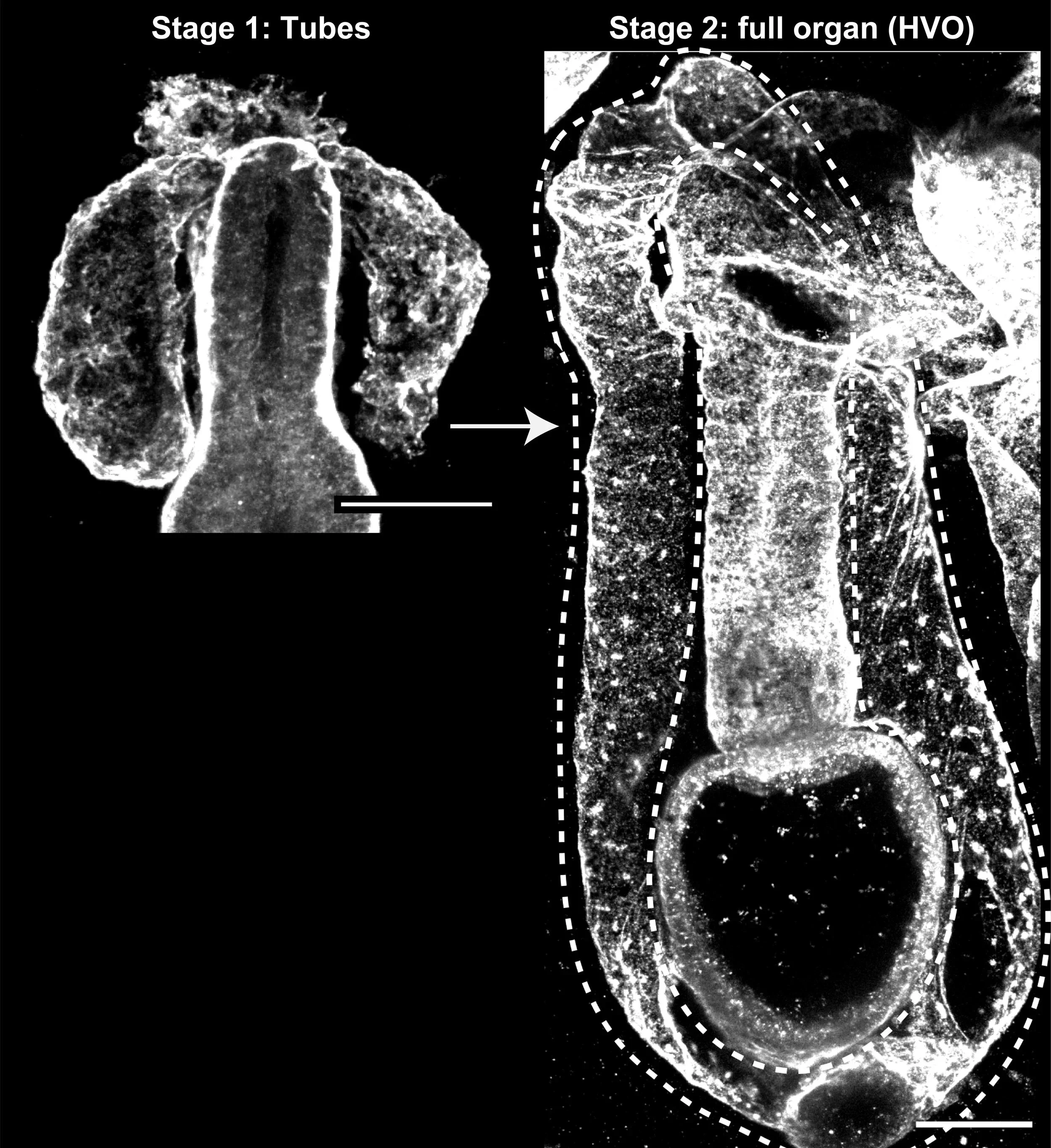
HVO highlighted by dotted lines, laminin (white). From Perillo et al., 2023
Mechanisms of tube orientation and shape
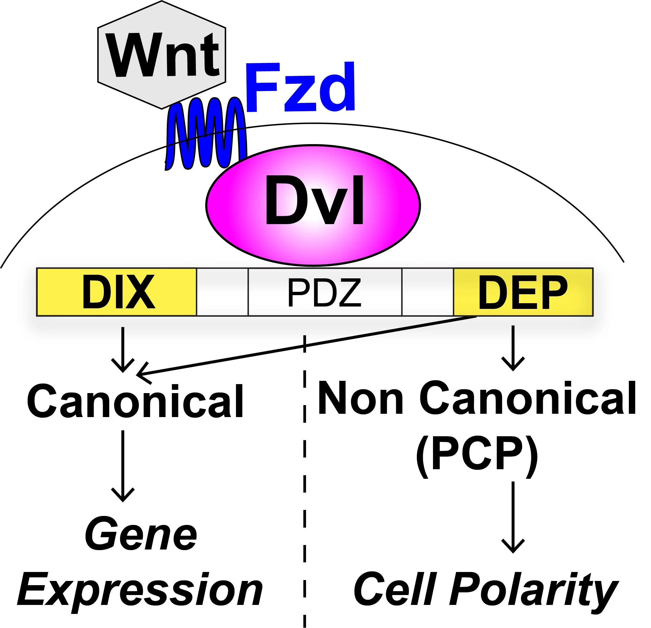
Formation of hollow tubes with a defined shape is a key developmental process that if compromised leads to malfunction of the entire organ. However, a general mechanism that explains the molecular basis for tube orientation and shape is still missing. We are exploring how Dishevelled (Dvl), a critical node that integrates the canonical and planar cell polarity (PCP) branches of the Wnt pathway, regulates tube orientation and shape.
This project is funded by a NIH/NIGMS MIRA ESI GRANT
Epithelial or migratory state? Mechanisms of organ integrity
To ensure organ integrity, sheets of polarized cells in an epithelial state create a barrier between the organ lumen and the outside. In pathological states, such as tissue fibrosis, cancer and aging, cells lose their epithelial state to become migratory through the epithelial-to-mesenchymal transition (EMT). We found that EMT can be pathologically induced in the HVO. In this project we will define the transcriptional signature of cells undergoing EMT.
This project is funded by a NIH/NIGMS MIRA ESI GRANT
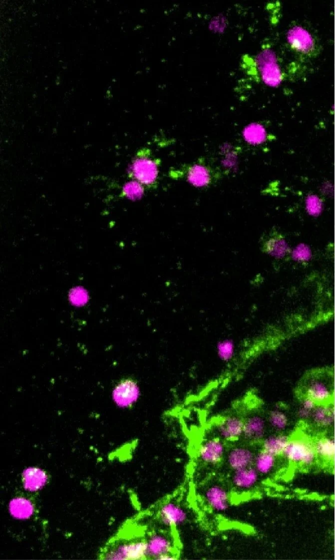
EMT from the HVO (nuclei in magenta, laminin in green)
Developmental mechanisms of organs specialized for environment sensing
Many marine animals develop through a planktonic larval stage that swims in the ocean. When conditions are optimal, larvae attach to a durable surface and metamorphose into adults. Because this process is irreversible, sensing of the substrate is critical to choose when and where to attach for settlement, and the consequent success of the animal. How do marine invertebrates, like the sea star, sense their environment to activate the process of metamorphosis? We are investigating how sea stars develop specialized sensing organs to link environment sensing to the decision of settlement.
This project is partially funded by the Company of Biologists ECR Visiting Fellowship award.
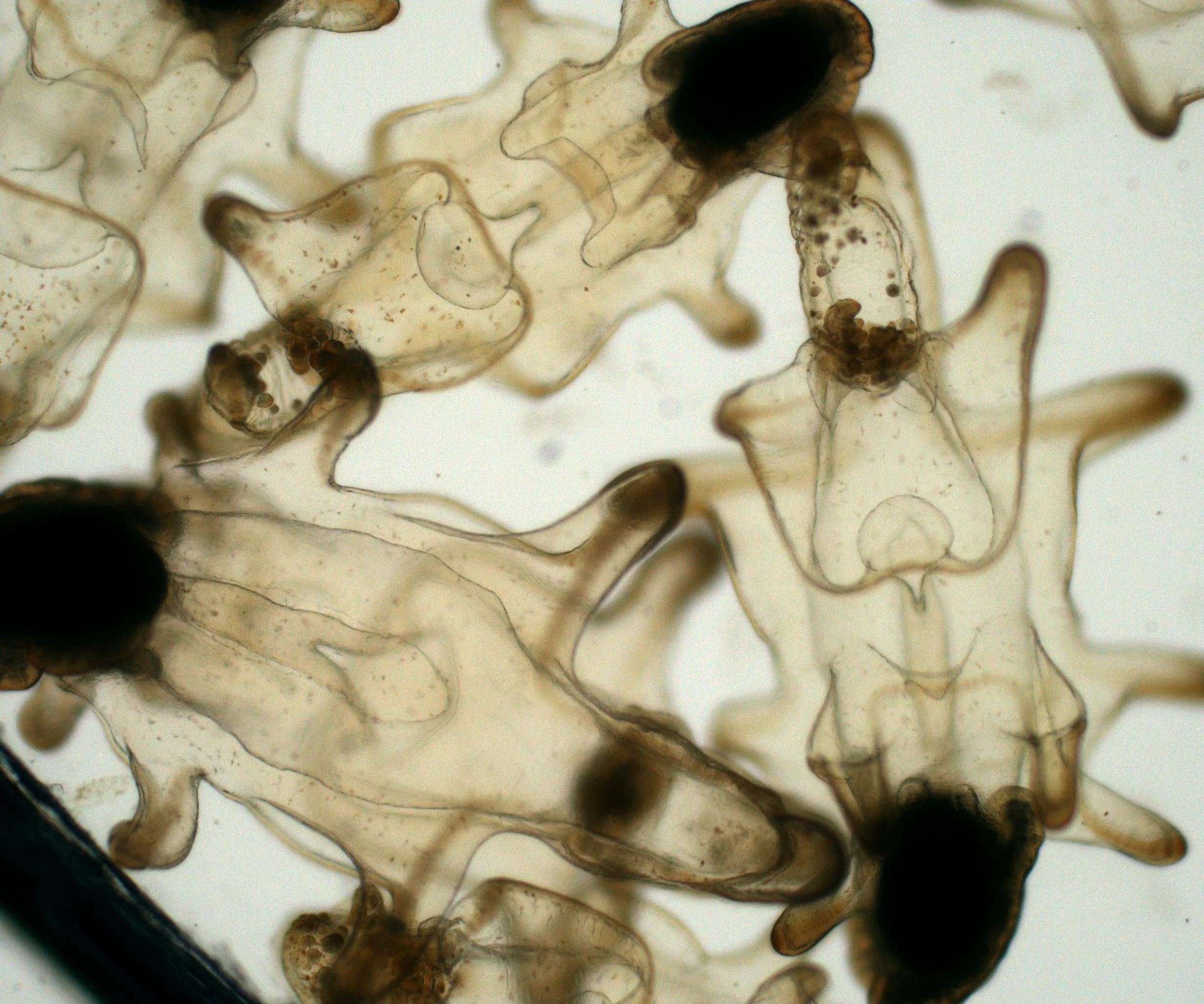
Sea star larvae ready for metamorphosis